Figure 2.
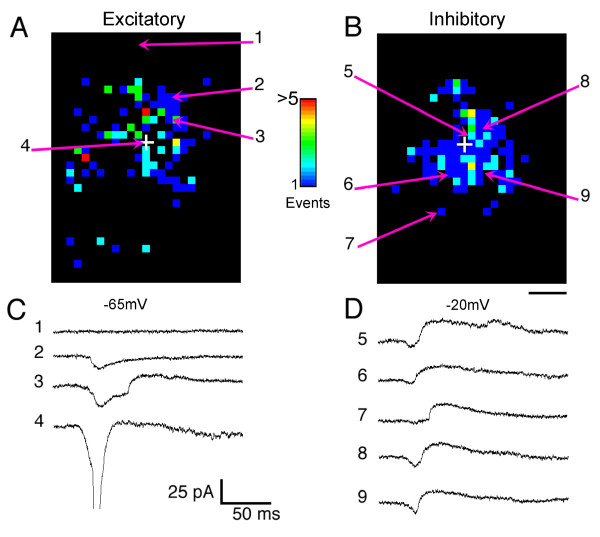
Maps of inhibitory and excitatory inputs are distinct. (A) Map of the locations and number of excitatory inputs to a pyramidal neuron (P58) evoked by photostimulation in a tangential slice of ferret visual cortex. Excitatory inputs arise from sites local (>300 μm) to and at longer distances from the cell body (indicated by the white cross). (B) Map of inhibitory inputs to the same neuron. Inhibitory inputs arise primarily within 300 μm of the cell body. (C, D) Electrophysiological recordings from sites indicated by the numbered arrows in (A, B). Photostimulation at the cell body evoked an action potential (4). Stimulation of other sites evoked excitatory (2, 3), inhibitory (7), or both excitatory and inhibitory inputs (3, 5, 6, 8, 9). To obtain the maps shown here, 1,467 locations were stimulated, mapping an area of approximately 2.1 mm2. Scale bar = 250 μm.

