Figure 1.
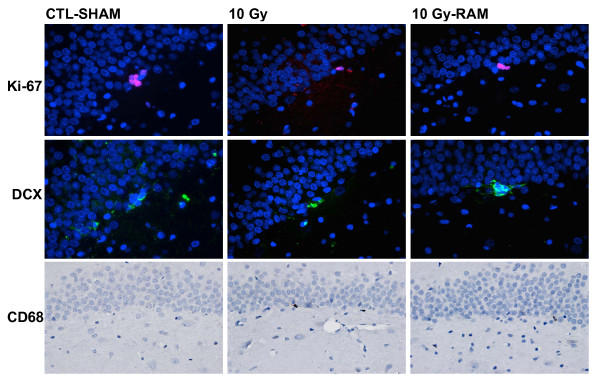
Representative images of immunohistochemical staining for Ki-67+ progenitors (red), DCX+ immature neurons (green), and CD68+ activated microglia (brown) in the SGZ and GCL, obtained at 400× from CLT-SHAM, 10 Gy, and 10 Gy-RAM group rats sacrificed at 12 weeks post-irradiation. Note the robust progenitor and immature neuron production, and sparse activated microglia, in CTL-SHAM tissue. Progenitor and immature neuron production are severely impaired, and activated microglia increased, in 10 Gy tissues. Impaired progenitor and immature neuron production are subtly but significantly improved in 10 Gy-RAM tissue.

