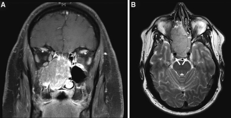
Fig. 2.
a and b Coronal and axial magnetic resonance images demonstrating a large SNUC mass filling both sides of the nasal cavity, bilateral ethmoid sinuses, and the right maxillary sinus in a 41-year-old man who presented with a 6-month history of nasal obstruction unresponsive to decongestants, steroids, or antibiotics. Arrows (part b) indicate carotid arteries with adjacent tumor involvement

