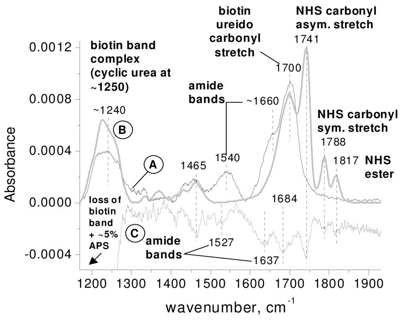
Figure 4.
Representative FTIR spectra of: (A) an APS surface exposed to biotin-NHS (for covalent attachment) and referenced to the pre-exposed APS surface; (B) a silicon oxide surface exposed to biotin-NHS (for physisorption) and referenced to the pre-exposed silicon oxide surface; and (C) a sonicated biotin-NHS treated APS surface referenced to the same biotinylated surface before sonication (amplified 5x).

