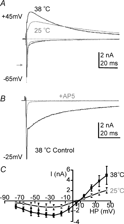
Figure 1. NMDAR-EPSCs double in amplitude on raising experimental temperature from RT to physiological.
A, the evoked EPSC at holding potentials of −65 mV or +45 mV for 37°C (black traces) and 25°C (grey traces) recorded from the same MNTB neuron. Zero current is indicated by the dashed line. The grey arrow indicates the peak fast EPSC at 25°C. B, superimposed traces from the same cell at a holding potential of −25 mV, before and during perfusion with the specific NMDAR antagonist d-AP5 (50 μm, grey trace). C, average current–voltage relationship measured at the peak of the slow EPSC shows the classic voltage dependence block at negative potentials. The potentiation with raised temperature is similar across all voltages, suggesting that the increase is not due to a change in magnesium sensitivity of the NMDAR. Data are means ±s.e.m. from 3 neurons per data point, each measured at both temperatures, from rat calyx of Held/MNTB.

