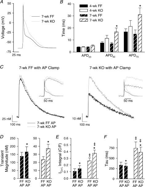
Figure 5. AP prolongation facilitated Ca2+ entry in 7-week KO.
A, representative AP recordings at 1 Hz. B, times to 20%, 50% and 70% repolarization (APD20, APD50 and APD70) (n= 16, 17, 11, 16; *P < 0.05 vs. corresponding FF). C, representative Ca2+ transients recorded during voltage-clamp stimulation with AP waveforms from A. Insets illustrate an expanded time scale with APs superimposed. Transient magnitude (D), integrated L-type Ca2+ current (E), and decay kinetics of the Ca2+ transient (F). (n for panels D, E and F: 7-week FF = 9, 8, 9; 7-week KO = 12, 4, 12; *P < 0.05 vs. equivalent FF AP, ‡P < 0.05 vs. equivalent 7-week FF).

