Figure 6.
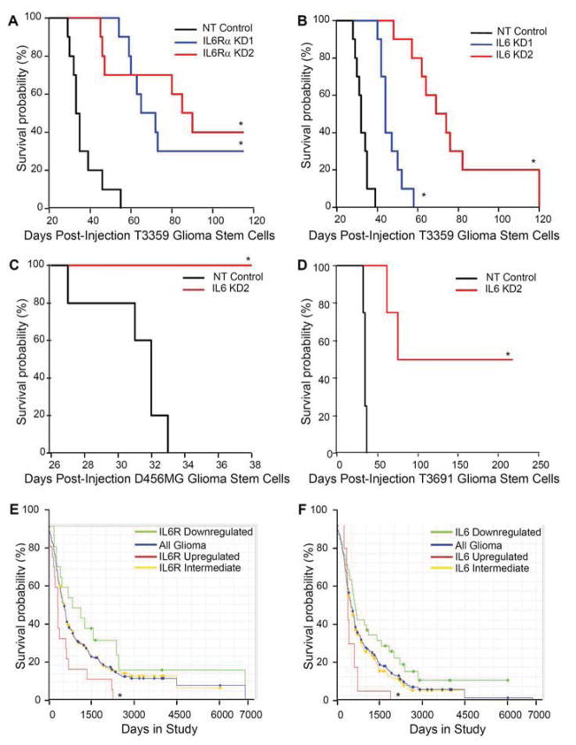
Targeting either IL6Rα or IL6 suppressed tumor growth and increased the survival of mice bearing intracranial xenografts. (A) Kaplan-Meier curves demonstrate increased survival with knockdown of IL6Rα in T3359 GSCs. 5000 GSCs infected for 24 hours with lentivirus expressing non-targeting control shRNA (NT) or two different shRNAs directed against IL6Rα (IL6Rα KD1 and IL6Rα KD2) were injected into the right frontal lobes of immunocompromised mice. *, p < 0.001 with comparison to non-targeting control. (B) Kaplan-Meier curves demonstrated increased survival with knockdown of IL6 (IL6 KD1 and IL6 KD2) compared to non-targeting control shRNA (NT) in T3359 GSCs injected as in A. *, p < 0.01 with comparison to non-targeting control. (C) Kaplan-Meier curves demonstrated increased survival with knockdown of IL6 when 10000 D456MG GSCs were treated an injected as in B. *, p < 0.001 with comparison to non-targeting control. (D) Kaplan-Meier curves demonstrated increased survival with knockdown of IL6 in T3691 GSCs injected as in B. p < 0.001 with comparison to non-targeting control. (E) Clinical data from the National Cancer Institute’s Repository for Molecular Brain Neoplasia Data (REMBRANDT) database indicates that higher IL6Rα mRNA levels in gliomas correlate with poor patient survival. *, p = 0.0076 with comparison of survival probabilities for patients with up-regulated IL6R expression to those with down-regulated IL6Rα expression. (F) Clinical data from REMBRANDT indicates that higher IL6 mRNA levels in gliomas correlate with poor patient survival. *, p = 0.0342 with comparison of survival probabilities for patients with up-regulated IL6 expression to those with down-regulated IL6 expression.

