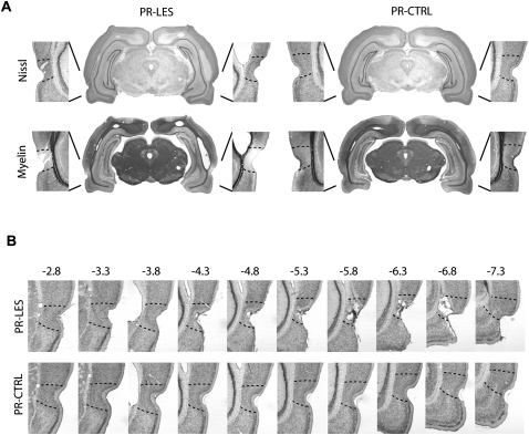
Figure 2.
Histological verifications of perirhinal cortical lesions. (A) Representative sections from the PR-LES and PR-CTRL groups. The upper panel shows Nissl-stained sections and the lower panel shows the adjacent sections stained for myelin. (B) Serial, Nissl-stained sections presented from anterior to posterior directions (from −2.8 to −7.3 mm from bregma).

