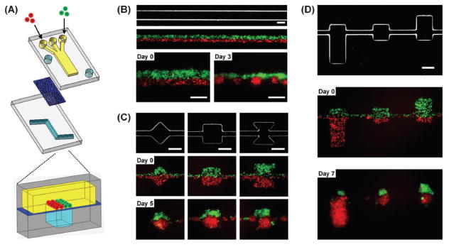
Fig. 1. Compartmentalized microfluidic system for cellular patterning.

(a) Schematic illustration of the microfluidic device. Two PDMS channel layers are separated by a semi-porous polycarbonate membrane which is rendered resistant to cell adhesion. The top channel is a straight channel (2 mm in width) with a dead-end. The bottom channel consists of a straight channel with or without chambers. Cells are introduced into the top channel using multiple laminar flows. (b–d) Micrographs of bottom layer geometry and actual cellular patterning. (b) Cellular patterning on a single straight bottom channel (200 μm in width). Two kinds of cells, MDA-MB-231 cells (green) and COS7 cells (red), are juxtaposed in the top layer as fluid focuses them together into one channel in the bottom layer. Each type of cell self-aggregates to form multiple spheroids. (c, d) Cellular patterning on the bottom channel with distinct geometric features to control either the shape (c) or size (d) of individual spheroids. MDA-MB-231 cells were stably transfected with EGFP and COS7 cells were labeled with CellTracker red. Scale bars: 200 μm.
