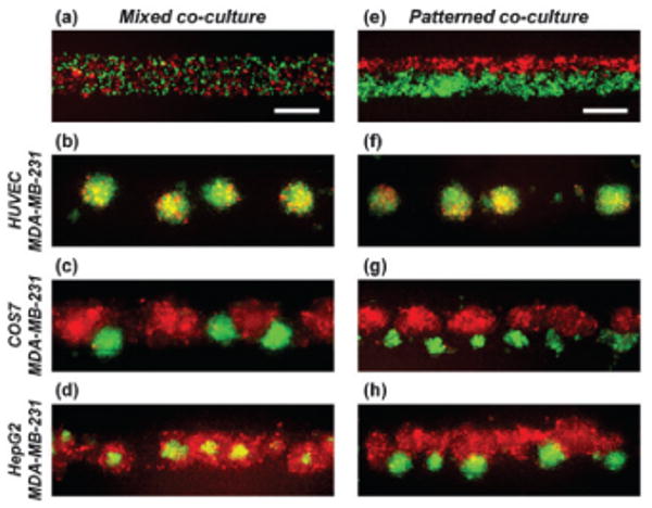
Fig. 4.

Formation of heterogeneous co-culture spheroids. Fluorescent images of mixed co-culture (a–d) and patterned co-culture (e–h) of two kinds of cells after 7 days in culture. MDA-MB-231 cells were co-cultured with either HUVECs (b, f), COS7 cells (c, g), or HepG2 cells (d, h). MDA-MB-231 cells were stably transfected with EGFP and all the other cells were labeled with CellTracker red. The bottom channel is a straight channel with a width of 200 μm. Scale bars: 200 μm.
