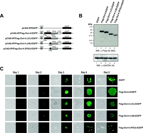
Figure 2. Different abilities of the Oct-4 deletion mutants to confer self-renewal.
(A) Schematic representation of the Oct-4–EGFP fusion construct and its derivatives with deleted domains. The functional domains located within Oct-4 are indicated as NTD (N-terminal domain), POU (POU DNA-binding domain) and CTD (C-terminal domain). (B) Immunoblot analysis of the expression of Oct-4 deletion mutants in stably transfected ZHBTc4 cells. ZHBTc4 ES cells were stably transfected with pCAG-IP/EGFP (labelled as EGFP; lane 1), pCAG-IP/FLAG-Oct-4–EGFP (Flag-Oct-4-EGFP; lane 2), pCAG-IP/FLAG-Oct-4(ΔC)–EGFP [Flag-Oct-4(ΔC)-EGFP; lane 3], pCAG-IP/FLAG-Oct-4(ΔN)–EGFP [Flag-Oct-4(ΔN)-EGFP; lane 4] or pCAG-IP/FLAG-Oct-4(POU)–EGFP [Flag-Oct-4(POU)-EGFP; lane 5], and total cell lysates were fractionated by SDS/PAGE (12% gels) and visualized by Western blotting with anti-FLAG (M2; Sigma) or anti-GAPDH (V-18; Santa Cruz Biotechnology) antibodies. The position of the prestained molecular-mass marker (New England Biolabs) is indicated to the left of the gel (molecular mass in kDa). (C) Self-renewal potentials of Oct-4 deletion mutants. ZHBTc4 ES cells stably transfected with pCAG-IP/EGFP (EGFP), pCAG-IP/FLAG-Oct-4–EGFP (Flag-Oct-4-EGFP), pCAG-IP/FLAG-Oct-4(ΔC)–EGFP [Flag-Oct-4(ΔC)-EGFP], pCAG-IP/FLAG-Oct-4(ΔN)–EGFP [Flag-Oct-4(ΔN)EGFP] or pCAG-IP/FLAG-Oct-4(POU)–EGFP [Flag-Oct-4(POU)-EGFP] were cultured in the presence of doxycycline. Phase-contrast (left-hand panels, grey colour) and fluorescence views (right-hand panels, green colour) are shown. Ab, antibody; WB, Western blot.

