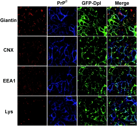
Figure 2. GFP–Dpl and PrPC localize in the Golgi apparatus.
Doubly transfected FRT cells were treated as in Figure 1(A) before being incubated with SAF-32 (against PrPC; blue) and primary polyclonal antibodies against different markers of intracellular compartments (CNX for the ER, giantin for Golgi and EEA1 for early endosomes; red). Secondary antibodies were Cy5-conjugated anti-(mouse Ig) antibody and TRITC-conjugated anti-(rabbit Ig) respectively. To label lysosomes, Lysotracker Red DND-99 (Lys, 1:10000 dilution in cell culture medium) was added to live cells 1 h before fixation and confocal imaging. Dpl was also visualized through the fluorescence of the GFP tag (green). Confocal microscopy was performed as described in Figure 1(A). The localization of GFP–Dpl and PrPC in the Golgi network is clearly evident from the merging of their respective signals with giantin. Scale bar, 10 μm.

