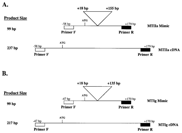
Figure 1.
Schematic representation of the MTIIa mimic cDNA (A). A 138-bp internal sequence (+18 to +155 bp from the start of translation) was deleted from the MTIIa cDNA to create the 99-bp MTIIa mimic cDNA. Shown for comparison is the full-length 237-bp MTIIa cDNA. Schematic representation of the MTIg mimic cDNA (B). A 118-bp internal sequence (+18 to +135 base pairs, from the start of translation) was deleted from the MTIg cDNA to create the 99-bp MTIg mimic cDNA. Shown for comparison is the full-length 217-bp MTIg cDNA. Indicated are the primer binding sites.

