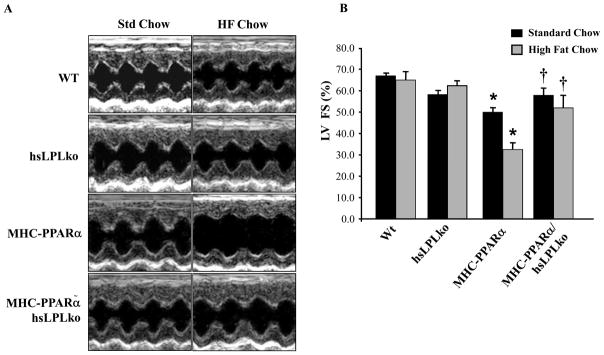
Figure 1. LpL-deficiency rescues ventricular dysfunction in MHC-PPARα mice.
A) Representative M-mode echocardiographic images of the LV from each genotype at baseline (one month) and after 4 weeks of HF diet. B) Bars represent mean (±SE) percent LV fractional shortening (FS), as determined by echocardiographic analyses. *p<0.05 vs WT and hsLpLko, †p<0.05 vs. MHC-PPARα mice on matched diet (n=6–8/group).

