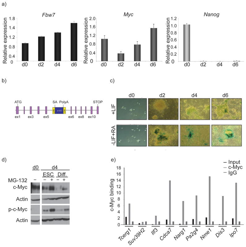
Figure 7. Patterns of Fbw7 and c-Myc expression in mouse embryonic stem cells.
a) mRNA transcript expression levels of Fbw7 (black), Myc (dark grey) and Nanog (light grey) in self-renewing (+LIF) and differentiating (−LIF, +RA) mouse ESC. Different days of differentiation (d0–6) are shown), b) Schematic diagram of Fbw7 gene-trap cassette. c) LacZ staining in Murine ESCs containing the Fbw7 gene trap cassette depicting upregulation of Fbw7 expression as Murine ESCs differentiate via the removal of LIF and the addition of Retinoic Acid (RA). d) Western blot showing stabilization of both phospho- (T58) and total-c-Myc in self-renewing and differentiating Murine ESCs treated with 20uM of proteosome inhibitor MG-132. e) ChIP assay was carried out using the specific regulators shown in Fig 6 revealing substantial enrichment upon c-Myc immunoprecipitation in Murine ESCs. Error bars indicate standard deviation (std) from 3 independent experiments.

