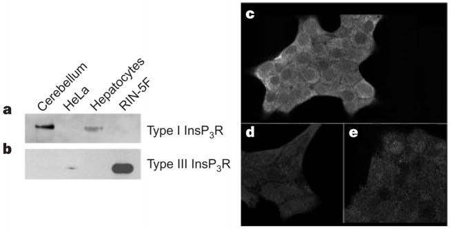
Figure 1.
RIN-5F cells preferentially express Type III InsP3R. a, b, Western blots were probed for types I and III InsP3R (a and b, respectively). Dog cerebellum and rat hepatocytes were positive controls for type I InsP3R; HeLa cells were a positive control for type III InsP3R. Lanes were loaded with 30 μg dog cerebellar microsomes (lane 1), 10 μg HeLa cell lysate (lane 2), 30 μg hepatic microsomes (lane 3), and either 30 μg (a) or 5 μg (b) of RIN-5F microsomes (lane 4). c, e, Cellular distribution of type III (c) and type I (e) InsP3R in RIN-5F cells. d, Nonspecific binding.

