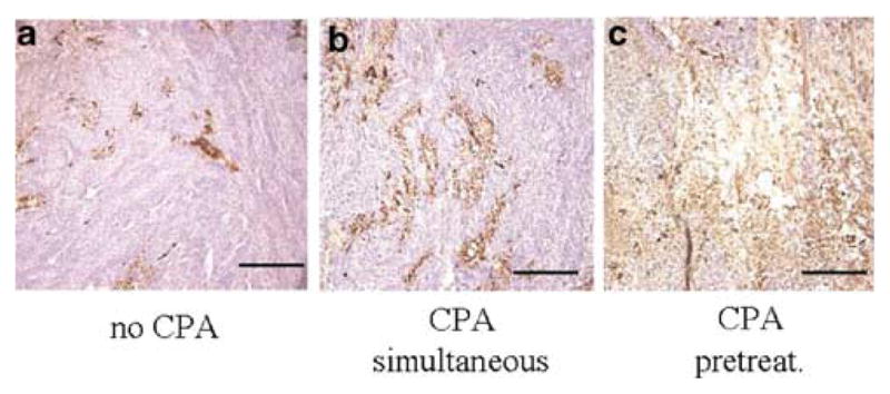
Figure 2.

Immunohistochemical detection of HSV capsid protein. In parallel to the experiment described in Figure 1, brain sections were also immunocytochemically processed for the expression of HSV1 capsid antigens. In panel a, a representative tumor section from animals treated only with hrR3 is shown with brown precipitates indicative of HSV capsid antigen expression. In panel b, a representative section is shown from tumors harvested from animals treated with hrR3 and CPA on the same day. In panel c, a representative section is shown from tumors harvested from animals treated with hrR3 and pretreated with CPA (2 days before). The scale bar represents 100 μm.
