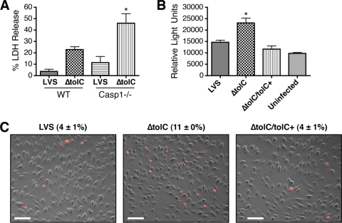
FIG. 5.
Hypercytotoxicity of the LVS ΔtolC is independent of caspase-1 and involves caspase-3. (A) muBMDM isolated from wild-type or caspase-1-deficient C57BL/6 mice were infected with the LVS or ΔtolC mutant at an MOI of 50. Cytotoxicity was quantified by measuring LDH release at 24 h p.i. The ΔtolC mutant caused significantly increased LDH release compared to that of the wild-type LVS for the caspase-1-deficient muBMDM (P < 0.05). Bars represent means ± SEM from three replicate samples. A representative experiment is shown. The experiment was repeated with similar results. (B) muBMDM isolated from C3H/HeN mice were uninfected or were infected with the LVS, ΔtolC mutant, or complemented strain (ΔtolC tolC+) at an MOI of 50. Activated caspase-3/-7 was measured at 17 h p.i. using a luminescence-based assay. Infection with the ΔtolC mutant caused a significant increase in caspase-3/-7 activity compared to that of the wild-type LVS (P < 0.05). Bars represent means ± SEM from three replicate samples. A representative experiment is shown. The experiment was repeated twice more with similar results. (C) muBMDM from C3H/HeN mice were infected as described for panel B and probed with an antibody against mature caspase-3 followed by a TRITC-conjugated secondary antibody. The images show overlays of caspase-3-positive cells (red) and corresponding phase-contrast images. The percentages of caspase-3-positive cells ± SEM were calculated from 10 separate fields and represent the averages from three independent experiments. The ΔtolC mutant caused significantly increased staining compared to that of the wild-type LVS (P < 0.05). Bars = 50 μm.

