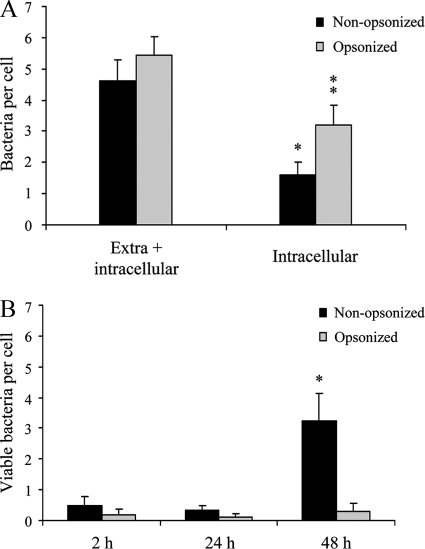
FIG. 2.
Phagocytosis and survival of B. pertussis in human macrophages. (A) GFP-expressing B. pertussis cells were incubated with human macrophages (MOI, 20) for 20 min at 37°C. After being washed, the cells were fixed and extracellular and intracellular bacteria were quantified by double immunofluorescence staining. The data represent the mean ± SD of three independent experiments. The number of intracellular IgG-opsonized B. pertussis bacteria was significantly different from the number of intracellular nonopsonized B. pertussis bacteria (*, P < 0.05). (B) GFP-expressing B. pertussis was incubated with human macrophages (MOI, 20) for 20 min at 37°C. After three washing steps, the cells were incubated with polymyxin B to kill extracellular bacteria and the number of CFU of B. pertussis per cell was determined at different time points postinfection. The data represent the mean ± SD of three independent experiments. The number of viable intracellular nonopsonized bacteria per cell at 48 h postinfection was significantly different from the number of viable intracellular bacteria per cell at both 2 and 24 h postinfection. (*, P < 0.05).

