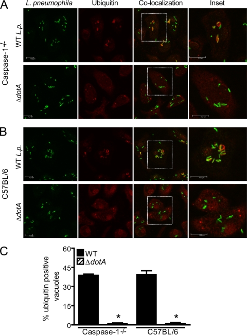
FIG. 4.
Pore formation is not a result of membrane damage by the Dot/Icm apparatus. BMMs from caspase-1−/− and C57BL/6 mice were infected with wild-type (WT) L. pneumophila (WT L.p.) or isogenic ΔdotA mutants for 1 h at an MOI of 10 in the presence of anti-L. pneumophila antibody. Recruitment of ubiquitin (red) to L. pneumophila vacuoles (green) in caspase-1−/− (A) or C57BL/6 (B) infected BMMs was visualized by confocal microscopy. Confocal high-resolution detail corresponds to ubiquitin recruitment to the vacuoles in the white inset. Scale bar, 10 μm. (C) Percentage of infected cells showing ubiquitin-positive L. pneumophila vacuoles upon infection with WT L. pneumophila or ΔdotA mutants. Data are representative of three independent experiments. *, P < 0.05 in comparison to BMMs infected with WT L. pneumophila.

