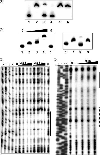
FIG. 5.
(A) Competition EMSA experiments with the hmbR promoter and MisR. A 326-bp hmbR fragment (YT173-hmbR-pLR) was end labeled with [γ-32P]ATP using T4 kinase and incubated with the MisR protein as described previously (52). Lane 1, DNA probe; lanes 2 to 6, DNA probe with MisR protein (77 pmol, 5 μM) (lane 2, no DNA competitor; lanes 3 and 4, probe with 1.5 and 2 μg specific DNA competitor, respectively; lanes 5 and 6, probe with 1.5 and 2 μg nonspecific DNA competitor, respectively). (B) (Left panel) Dose-dependent shift of the hemO promoter with MisR. Lanes 1 and 5, free probe; lanes 2 to 4, 0.6, 2.6, and 3.8 μM MisR protein, respectively. (Right panel) Competition EMSA with the hemO promoter. Lane 6, free probe; lanes 7 to 9, DNA probe with MisR protein (38 pmol, 2.5 μM). One microgram of cold specific DNA was added to lane 8, while 1 μg of nonspecific DNA was added to lane 9. (C and D) DNase I protection assays with the hemO promoter (C) and the hmbR promoter (D). The noncoding strand of the promoter fragment was end labeled with 32P as described in Materials and Methods, incubated with increasing amounts of MisR for 20 min at 30°C, and then subjected to DNase I digestion in a 20-μl (total volume) reaction mixture. MisR∼P was prepared by incubation with acetyl phosphate (50 mM) at 37°C for 30 min. For hemO, the amounts of MisR tested were 0, 93, 186, 232, and 278 pmol and the total amounts of MisR∼P tested were 0, 93, 139, 232, and 371 pmol. For hmbR, the amounts of MisR tested were 0, 170, 340, 510, and 850 pmol. The results for dideoxy chain termination sequencing ladders corresponding to the probes are shown on the left (lanes A, T, G, and C). The solid line indicates the sequence protected by MisR, while the Fur protected region determined by Delany et al. (10) is indicated by a dotted line.

