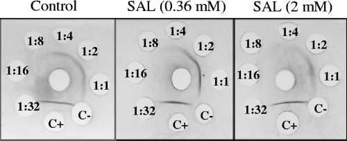
FIG. 4.
Immunodiffusion analysis of CP5 extracts. Thirty microliters of monospecific antiserum to serotype 5 was added to the center well. Thirty-microliter samples of undiluted and twofold serially diluted (indicated inside the well) CP5 extracts of Newman strain organisms, cultured or not with different concentrations of SAL, were added to the outer wells. Precipitin lines were visualized after staining with Coomassie brilliant blue. C+, positive control [undiluted extract of S. aureus Reynolds (CP5) strain]; C−, negative control [undiluted extract of S. aureus Reynolds (CP−) strain]. Precipitin lines were visible up to a 1:4 dilution of capsular extracts in the control without SAL, up to a 1:2 dilution at a 0.36 mM SAL concentration, and at only a 1:1 dilution at the highest SAL concentration (2 mM).

