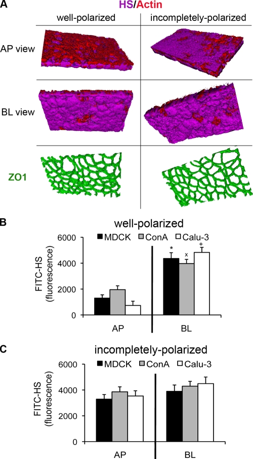
FIG. 2.
HSPGs are expressed on the AP surface in incompletely polarized cells. In well-polarized monolayers grown on Transwell filters, there was a greater amount of HSPGs on the BL surface than on the AP surface. In incompletely polarized monolayers, there was increased expression of HSPGs on the AP surface. (A) 3D reconstruction of z-stack images acquired by confocal microscopy. Shown are AP and BL projections of well-polarized and incompletely polarized Calu-3 cells stained with an antibody against the HS chain of HSPGs (FITC-HS; purple), with phalloidin (red) to visualize actin, and with ZO-1 (green) to visualize tight junctions. (B and C) FITC-HS fluorescence (in arbitrary units) measured in a fluorescence plate reader. Well-polarized (B) or incompletely polarized (C) MDCK, ConAr, and Calu-3 cells were stained with FITC-HS added to the AP or BL surface. Shown are the means ± SD for three separate experiments. *, P < 0.05 compared to AP surface-stained MDCK cells; ×, P < 0.05 compared to AP surface-stained ConAr cells; +, P < 0.05 compared to AP surface-stained Calu-3 cells.

