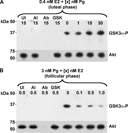
FIG. 4.
Increasing Pg promotes Akt kinase activity. Pex cells were incubated in E2/Pg concentrations reflective of the menstrual cycle luteal (A) or follicular (B) phase and then challenged with N. gonorrhoeae strain 1291, as noted, for 3 h. Akt was immunocaptured from pex cell lysates, and the kinase activity was assessed as described in the text by using a GSK-3α peptide substrate. The presence of an ∼37-kDa band, after immunoblotting with the phospho-GSK-3α/β (Ser21/9) (rabbit) antibody, is consistent with phosphorylation of the GSK-3α peptide (GSK-3α-P) and is indicative of Akt activity. The membranes were stripped and reprobed using the mouse anti-Akt antibody B-1 to ensure the presence of comparable amounts of Akt kinase under each assay condition (lower image in panels A and B). The hormone concentrations (0.4 nM E2 plus 0 to 30 nM Pg in panel A; 3 nM Pg plus 0 to 1 nM E2 in panel B) used are noted across the top of each membrane image. UI, uninfected pex cells; AI, Akt inhibitor VII was included in the assay; Ab, the agarose-conjugated anti-Akt capture antibody, C-20AC, was omitted from the initial capture step; GSK, the GSK-3α peptide substrate was omitted from the kinase reaction.

