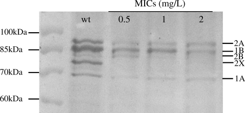
FIG. 1.
PBP profiles of S. uberis ATCC 19436 wild type and its derivatives, presenting increasing MICs. Proteins were separated by SDS-PAGE, and Bocillin-PBP complexes were visualized by fluorography. Molecular mass markers are indicated at the left of the fluorogram, and PBP numbers, named after their homologues in S. pneumoniae, are at the right. The wild type (wt) and cycled mutants with increased penicillin MICs are indicated at the top.

