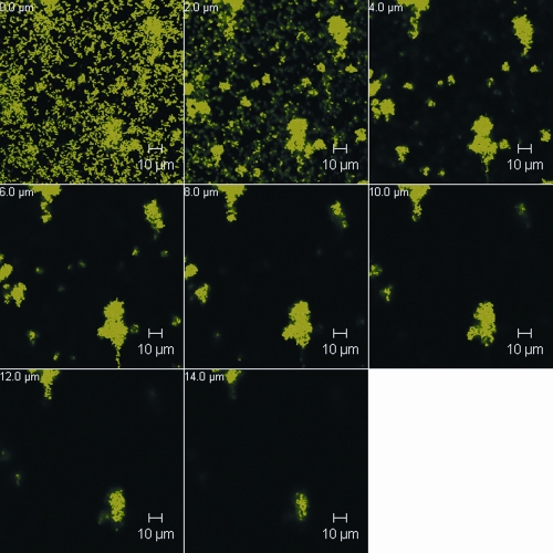
FIG. 2.
CLSM images of a representative section of a P. gingivalis ATCC 33277 18-h biofilm grown in a flow cell and stained with BacLight. Horizontal (x-y) optodigital sections, each 2 μm thick over the entire thickness of the biofilm (z), were imaged using a 63× objective at 512 by 512 pixels (0.28 μm per pixel), with each frame at 143.86 μm (x) by 143.86 μm (y).

