FIG. 1.
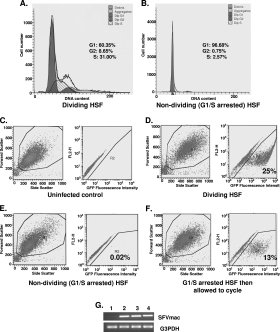
Transduction and transgene expression efficiency by SFVmac in cycling and G1/S phase-arrested HSF cells. Cell cycle analysis results in cycling and arrested cells are shown in panels A and B, respectively. The populations of cells at different phases of the cell cycle are given as a percentage of the total population. Cells in G1/S phase were at a 97% level. FACS analyses were performed at the same time for GFP expression. (C) FACS results for GFP expression in uninfected dividing cells. (D and E) FACS results of dividing and growth-arrested GFP-expressing cells, respectively, infected with SFVmac vector. (F) GFP expression in cells where medium containing aphidicolin was removed from the culture and replaced with fresh medium without aphidicolin, allowing cells to undergo cell division. (G) DNA amplified by PCR from SFVmac vector-transduced HSF cells. Lane 1 is mock transduced cells, lane 2 is cycling HSF cells transduced with SFVmac vector, and lane 3 represents G1/S phase-arrested cells transduced with foamy vector. PCR results from growth-arrested cells infected with foamy virus vector and released into cycle 24 h later are shown in lane 4. DNA was amplified using pol-specific forward 5′-TGTAATACCACTCCAAGCCTGGAT-3′ and reverse 5′-GACTTTCAGAAAAGTAGCGTCTCG-3′ primers.

