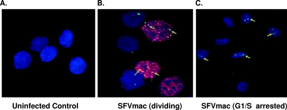
FIG. 2.
SFVmac genome localization in dividing and growth-arrested cells. For confocal microscopy cells were labeled with BrdU (red) to distinguish between dividing and growth-arrested cells. SFV-1 genome was identified with in situ hybridization using gag-specific probe (green). The nucleus was counterstained with DAPI (blue). (A) In situ staining of uninfected control cells. (B and C) Dividing and growth-arrested cells, respectively, transduced with foamy virus vector.

