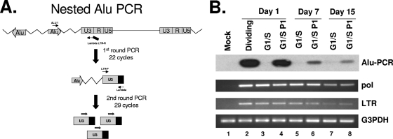
FIG. 3.
Alu PCR of genomic DNA isolated from SFVmac vector-infected dividing or growth-arrested cells. (A) Schematic representation of an Alu PCR methodology. DNA was copied using an Alu sequence-specific primer and amplified with an SFVmac LTR-specific primer tagged with a lambda sequence. (B) Alu PCR with samples isolated from dividing and growth-arrested cells (top panel). The Alu PCR-amplified products were hybridized to radioactive [γ-32P]ATP-labeled LTR-specific probe. Lane 1 (mock) contains PCR results from uninfected control cells. Lane 2 (dividing) is the PCR product with DNA isolated from SFVmac-transduced dividing cells 24 h after transduction. Lanes 3, 5, and 7 represent PCR results from G1/S-arrested cells harvested at days 1, 7, and 15 posttransduction, respectively. Lanes 4, 6, and 8 are PCR products for cells that were arrested in G1/S for 1, 7, and 15 days, respectively, and then allowed to resume a normal cycle for 72 h. The second and third panels represent corresponding semiquantitative regular PCR detecting the pol and LTR region of SFVmac. For level of sample recovery, the DNAs were amplified with primers specific to G3PDH (glyceraldehyde-3-phosphate dehydrogenase) (bottom panel).

