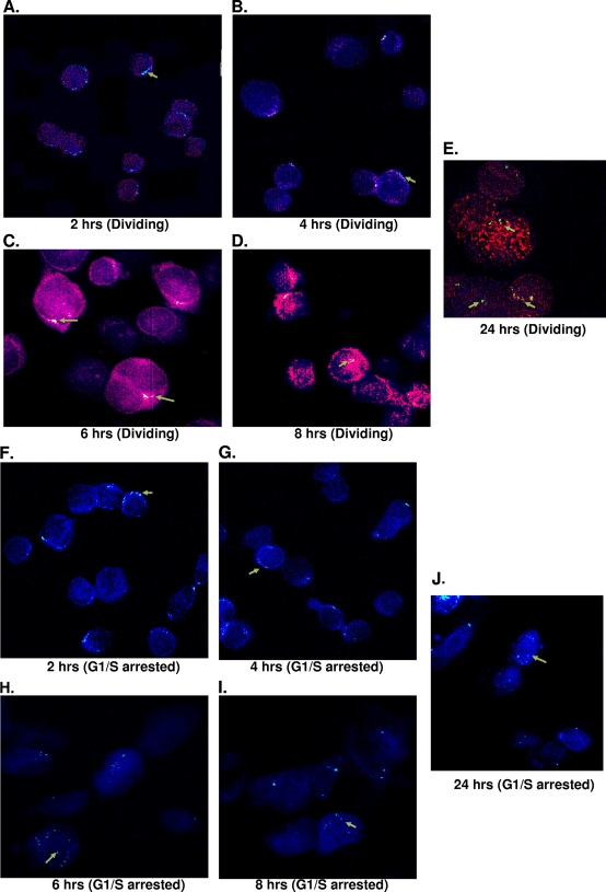
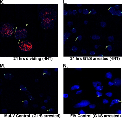
FIG. 5.
SFVmac genome localization in the nuclei of nucleoporin labeled dividing and growth-arrested cells at different time points after infection with SFVmac vector. BrdU (red)-labeled cells were permeabilized, and in situ hybridization was performed by probing for SFVmac genome (green). The nucleus was defined by immunostaining with antibody to nucleoporin, Nup153 (blue). (A to E) In situ hybridization for foamy virus genome with immunolabeling for Nup153 at the indicated time points in dividing cells. (F to J) Genome localization at different time points in cells arrested at the G1/S phase. Foamy virus genome localization in dividing and growth-arrested cells infected with integrase defective vector at 24 h posttransduction is shown in panels K and L, respectively. In situ hybridization for MuLV and FIV controls at 24 h posttransduction are represented in panels M and N, respectively.


