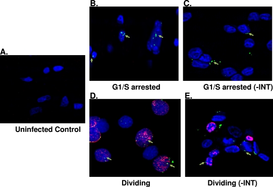
FIG. 6.
SFV Gag localization in the absence of integrase. Dividing and nondividing cells were transduced with SFVmac vector defective for integrase, and immunohistochemistry was performed with antibody against Gag (green) and nucleoporin with anti-Nup153 (blue). BrdU labeling is represented by a red stain showing dividing cells. (A) Uninfected control. (B and C) Growth-arrested cells transduced with wild-type SFVmac vector and integrase defective vector, respectively. Gag localization in dividing cells are represented in panels D and E. “-INT” indicates integrase-defective SFVmac vector.

