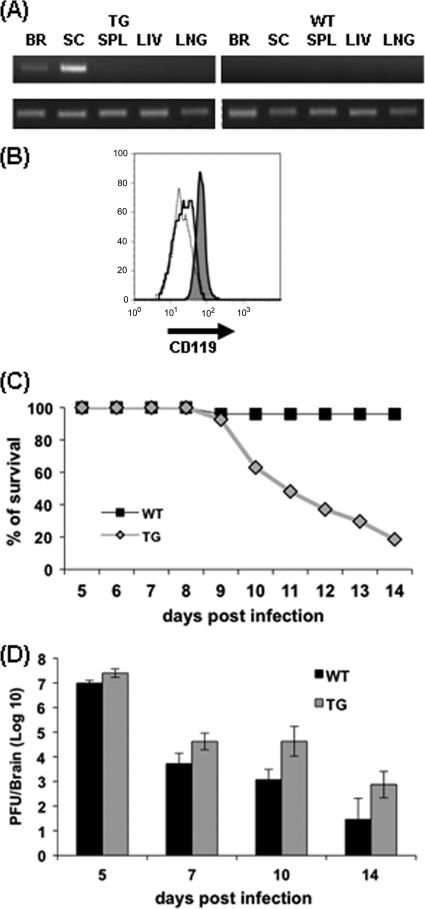
FIG. 1.
IFN-γ signaling in oligodendrocytes protects BALB/c mice. (A) Expression of the dnIFN-γR1 transgene mRNA in the CNS. RNA obtained from brain (BR), spinal cord (SC), spleen (SPL), liver (LIV), and lungs (LNG) of naïve homozygous TG and WT BALB/c mice, amplified by PCR by using transgene (top)- and hypoxanthine phosphoribosyltransferase (HPRT) (bottom)-specific primers as previously described (10). (B) Surface expression of IFN-γR1 (CD119; PharMingen, San Diego, CA) on oligodendrocytes obtained from spinal cords of naïve TG and WT animals by trypsin digestion and enrichment on 30%/70% Percoll (Pharmacia, Uppsala, Sweden) gradients as described previously (11, 17). Flow cytometry histogram of CD119 expression on CD45− O4+ oligodendrocytes from TG mice (dark gray) and WT mice (solid line). The dotted line represents isotype control staining. (C) Survival rates of TG and WT mice following JHMV infection. (D) Virus replication in brains of TG and WT mice determined by plaque assay of tissue homogenates on DBT cell monolayers. Each time point represents the average of ≥3 individuals ± the standard deviation. At all time points p.i., differences between TG and WT mice were statistically significant (P < 0.05).

