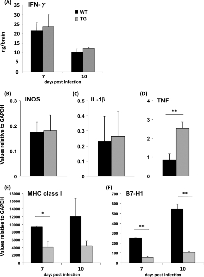
FIG. 4.
Altered gene expression by impaired IFN-γ signaling in oligodendrocytes. (A) Homogenates of individual brains from infected TG and WT mice were used to determine IFN-γ levels by enzyme-linked immunosorbent assay as described previously (29). RNA was extracted from the brains of TG and WT mice at 10 days p.i. using the Trizol reagent. Expression of iNOS (B), IL-1β (C), and TNF mRNA (D) relative to that of GAPDH (glyceraldehyde-3-phosphate dehydrogenase) was determined by quantitative reverse transcription-PCR (qRT-PCR), using previously described primers (12, 17). Each time point represents the average for ≥3 individuals ± the standard deviation. (E) Expression of MHC class I and B7-H1 (F) mRNA determined by qRT-PCR as described previously (17) in CD45− O4+ oligodendrocytes purified from groups of six to seven mice at days 7 and 10 p.i. Expression levels were normalized to GAPDH by using the following formula:  ×1,000, where CT is the threshold cycle. Expression levels in naïve mice were subtracted. Statistically significant differences for TNF, MHC class I, and B7-H1 mRNA are denoted by a single asterisk (P < 0.05) or a double asterisk (P < 0.005).

