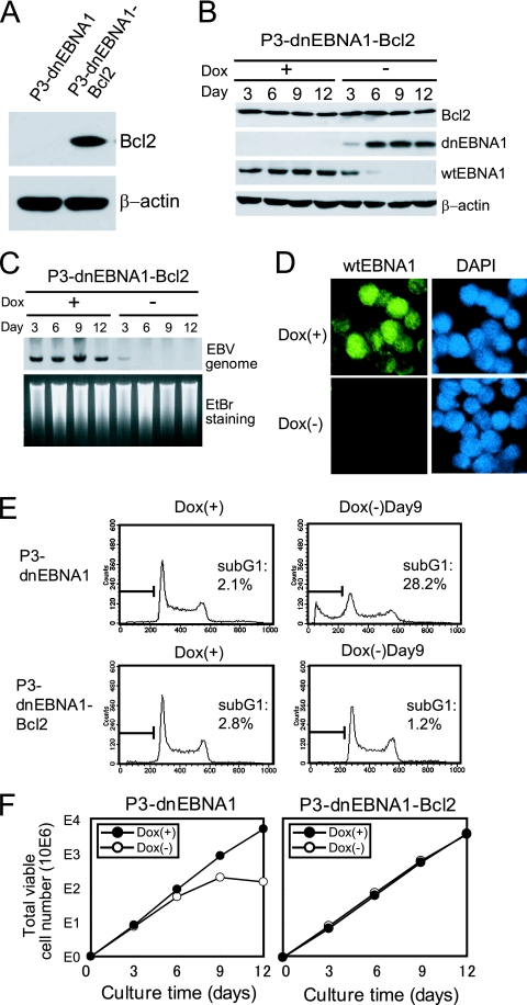
FIG. 4.
Stable overexpression of Bcl-2 rescues P3HR-1 cells from dnEBNA1-induced growth inhibition and apoptosis. (A) P3-dnEBNA1 and P3-dnEBNA1-Bcl2 cells were subjected to Western blot analysis with anti-Bcl-2 and anti-β-actin antibodies. (B) P3-dnEBNA1-Bcl2 cells cultured in the absence (−) or presence (+) of Dox were subjected to Western blot analysis with EBV-immune human serum (reactive to dnEBNA1 and wt EBNA1), anti-Bcl-2, and anti-β-actin antibodies. (C) P3-dnEBNA1-Bcl2 cells cultured in the absence (−) or presence (+) of Dox were subjected to Southern blot analysis to detect the EBV genome. (D) P3-dnEBNA1-Bcl2 cells were cultured in the absence (−) or presence (+) of Dox for 12 days. The cells were processed for immunofluorescence analyses with EBV-immune human serum (reactive to wt EBNA1, but not to dnEBNA1) as a primary antibody and FITC-conjugated anti-C3C antibody as a secondary antibody. (E) P3-dnEBNA1 cells and P3-dnEBNA1-Bcl2 cells were cultured in the absence (−) or presence (+) of Dox for 9 days. Cell cycle analysis was performed, and the percentages of cells in the sub-G1 fraction were determined. (F) P3-dnEBNA1 and P3-dnEBNA1-Bcl2 cells were cultured in the absence (−) or presence (+) of Dox. Viable cells were counted every 3 days, and the cells were split. The total numbers of viable cells derived from the initial cultures were calculated and plotted.

