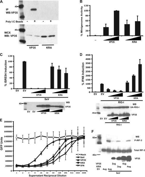
FIG. 2.
KRA mutant VP35 lacks dsRNA binding and IFN antagonist activity but retains polymerase cofactor function. (A) Wild-type but not KRA mutant VP35 binds to poly(I·C)-Sepharose beads. WT or KRA mutant VP35 transfected cell lysates were incubated with poly(I·C) beads (+) or control Sepharose beads (−). The precipitated material was analyzed by Western blotting for VP35 (upper panel). Whole-cell extracts (WCX) were probed for VP35 (lower panel). (B) Polymerase cofactor activity of WT and KRA mutant VP35 was assessed with an EBOV minigenome assay. Wedges represent relative amounts of transfected WT or mutant VP35 expression plasmid. The value obtained with the higher concentration of WT VP35 was set to 100%. (C) WT and KRA mutant VP35 inhibition of SeV-induced ISG54 promoter activation. Cells were transfected with empty vector (EV) or increasing amounts (indicated by wedges) of WT VP35 or KRA mutant VP35 expression plasmid, an ISG54 promoter-firefly luciferase reporter plasmid, and a constitutively expressed Renilla luciferase reporter plasmid. Firefly luciferase activity was normalized to Renilla luciferase activity, and the activities of the EV-transfected and SeV-infected samples were set to 100% (top panel). Western blots show expression levels of WT and KRA mutant VP35 (lower panel). (D) WT and KRA mutant VP35 inhibition of RIG-I-induced IFN-β-promoter activation. Cells were transfected with empty vector (EV) or increasing amounts (indicated by wedges) of WT or KRA mutant VP35 expression plasmid, an IFN-β-promoter-firefly luciferase reporter plasmid, and a constitutively expressed Renilla luciferase reporter plasmid. Uninduced samples received additional empty vector DNA, and induced samples (RIG-I) received HA-tagged RIG-I expression plasmid. Firefly luciferase activity was normalized to Renilla luciferase activity, and the activities of the EV, RIG-I-transfected samples were set to 100% (top panel). Western blots (WB) show expression levels of WT and KRA mutant VP35 (lower panel). (E) IFN bioassay to assess suppression of endogenous IFN-α/β production by WT and KRA mutant VP35. Cells were transfected with 4 μg of empty vector (○ and •), VP35 (▴), or KRA mutant VP35 (▪) expression plasmids. Cells were mock infected (open symbol) or infected with SeV (solid symbols). Two-fold dilutions of UV-irradiated supernatants were applied to Vero cells. Direct treatment of Vero cells with dilutions of human IFN-β were included to generate a standard curve (×). Treated Vero cells were then infected with NDV-GFP, and virus replication was quantified with a fluorescence plate reader. (F) Inhibition of SeV-induced IRF-3 (S396) phosphorylation by WT and KRA mutant VP35. Cells were transfected with indicated amounts of empty vector (EV), WT, or KRA mutant VP35. Cells were mock infected or infected with SeV. Lysates were analyzed by Western blotting using anti-phospho-S396 IRF-3 (P-IRF-3) antibody (top panel), total IRF-3 antibody (middle panel), and an anti-VP35 antibody (lower panel). Error bars represent one standard deviation of results of at least three independent experiments.

