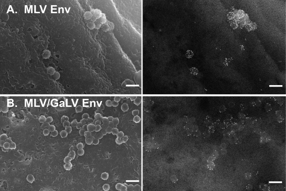
FIG. 2.
Distribution of MLV Env relative to HIV-1 assembly sites. 293T mCAT-1 cells were cotransfected with a plasmid expressing late domain-defective HIV-1 Gag and a plasmid expressing YFP-tagged MLV Env (A) or YFP-tagged MLV/GaLV Env (B). Env was labeled with 12-nm gold and imaged by SEM. Left, secondary electron images of HIV-1 assembly sites. Right, backscatter electron images of gold-labeled Env. Scale bars, 200 nm.

