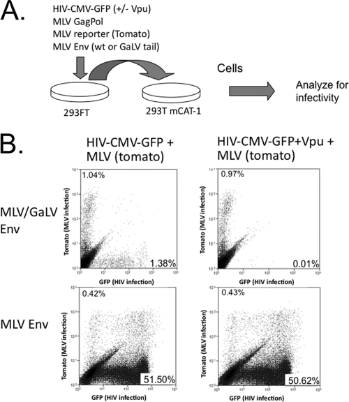
FIG. 7.
Vpu does not prevent the production of infectious MLV particles. (A) Schematic of dual infection assay. 293FT cells were transfected with HIV-1 and MLV assembly components, along with MLV or MLV/GaLV Env. At 48 h posttransfection the supernatant was transferred to 293T mCAT-1 cells. The ratio of HIV-1 to MLV was adjusted so that similar HIV-1 and MLV infectious particles were produced with MLV/GaLV Env and so that same ratio was used in each of the four transfections. (B) Flow cytometry output of 293T mCAT-1 infections. MLV infections display red fluorescence (y axis), and HIV-1 infections display green fluorescence (x axis). Infectivity is shown in each plot as percentage of the 293T mCAT-1 cells infected, excluding double-positive cells.

