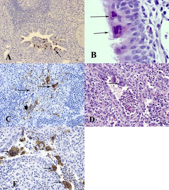
Figure 1.
Histopathological changes and the presence of viral antigen in pigs experimentally infected with Hendra virus. (A) Lung (minipig MP5): 5 dpi. Viral antigen was detected in epithelial cells of bronchioles (arrow). (B) Nasal Turbinate (minipig MP4): 7 dpi. Formation of epithelial syncytial cells (arrows). (C) Submandibular lymph node (minipig MP4): 7 dpi. Viral antigen was observed within reticular dendritic cells (arrows) as well as multinucleated syncytial cells (arrowhead). (D) Lung (Landrace pig P1): 5 dpi. A bronchiole shows attenuation and necrosis of the epithelium and formation of syncytial cells (arrow). (E) Lung (Landrace pig P1): 5 dpi. Positive immunostaining was observed within bronchiolar epithelial syncytial cells (arrows). (B, D) Hematoxylin and eosin (HE) stain; (A, C, E) immunohistochemistry/DAB/hematoxylin (IHC) staining. Bar = 20 μm. Magnification of B: ×100 objective (A color version of this figure is available at www.vetres.org).

