
Figure 1. MMP-9 Is Induced in BM Cells after BM Ablation.
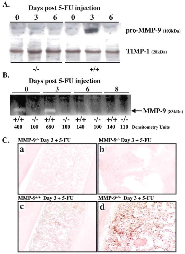
(A and B) MMP-9−/− and MMP-9+/+ mice received a single dose of 5-FU i.v. BM cells obtained at different time points after 5-FU injection were cultured in serum-free medium overnight. BM cell supernatants were assayed for pro-MMP-9 by Western blot (A) and active MMP-9 by gelatin zymography (B). Molecular weight (kDa) are shown (n = 5/group).
(C) Immunohistochemistry of BM sections three days after 5-FU injection for pro-MMP-9, which shows brown staining of stromal and hematopoietic elements in MMP-9+/+ mice (c and d), but not in MMP-9−/− mice (a and b); magnification ×100, (a and c) and ×400 (b and d).