
Figure 4. MMP-9 Mediated Release of sKitL Enhances Hematopoietic Reconstitution.
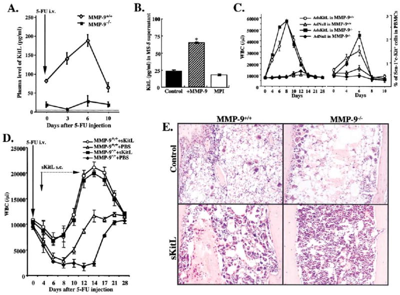
(A) MMP-9−/− and MMP-9+/+ mice were injected i.v. with a single dose of 5-FU and the plasma obtained from peripheral blood (PB) was assayed for sKitL by ELISA (p< 0.05, n = 6/group).
(B) Confluent MS-5 murine stromal cells, which express mKitL, were treated with recombinant active MMP-9 or the MPI CGS 27023A for 24 hr.*p< 0.001.
(C) MMP-9−/− and MMP-9+/+ mice were injected i.v. on day 0 with a single dose of Ad vector encoding for sKitL (AdsKitL) or no transgene (AdNull). PB was taken at indicated days (n = 6/group). Injection of AdsKitL resulted in sKitL plasma levels of 5399 ± 50 and 5126 ± 102 pg/ml on day 5 in MMP-9−/− and MMP-9+/+ mice, respectively. PBMCs were stained for Sca-1 and c-Kit and analyzed by FACS.
(D) MMP-9−/− and MMP-9+/+ mice were injected with recombinant sKitL from day 3–11 after 5-FU therapy (n = 10/group). WBC counts were determined at indicated time points.
(E) H&E staining of BM sections 4 days after 5-FU marrow suppression in MMP-9−/− and MMP-9+/+ mice without (control) and with sKitL Magnification × 200.