
Figure 6. Chemo/Cytokine-Induced HSC Mobilization Is Impaired in MMP-9−/− Mice.
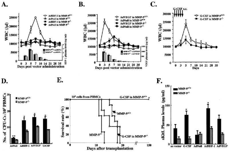
(A–C) MMP-9−/− and MMP-9+/+ mice were injected i.v. with a single dose of AdSDF-1, AdVEGF, and AdNull vector or s.c. with recombinant G-CSF from day 0–5 (n = 10 mice in each group). Elevated chemokine levels for SDF-1 and VEGF were achieved by adenoviral gene delivery of SDF-1 and VEGF ([A] and [B], bar graph insert). WBC counts were determined following AdSDF-1 (A), AdVEGF (B), and G-CSF treatment in MMP-9+/+ mice (C).
(D) Mobilized PBMCs were plated in a colony assay. The number of mobilized progenitor cells (CFU-C) was determined (n = 10, *0.05, **p< 0.01) on day 5 (AdSDF-1), on day 3 (AdVEGF), and on day 5 (G-CSF).
(E) PB of MMP-9−/− and MMP-9+/+ mice treated with or without G-CSF was obtained on day 5. PBMCs were transplanted into lethally irradiated syngeneic animals. Survival of transplanted recipients was monitored (n = 8/group, *p< 0.001).
(F) Plasma of MMP-9+/+ and MMP-9−/− mice 5 days after G-CSF, AdNull, AdSDF-1, and AdVEGF injection and untreated controls was assayed for sKitL (n = 6/group, *p< 0.03).