
Figure 3.
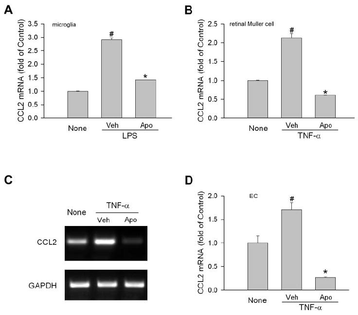
Involvement of NAD(P)H oxidase in CCL2 production in retinal cells. (A) Retinal microglial cells were pretreated with apocynin (Apo, 0.5 mM) or vehicle (Veh) for 0.5 hour Cells were then treated with LPS (30 ng/mL) for 3 hours. Total RNA was extracted, and CCL2 mRNA was measured by quantitative PCR. Cells without treatment (None) were used as control (n = 4). #P < 0.05 compared with None. *P < 0.05 compared with vehicle. (B) Retinal Müller cells (rMC-1) were pretreated with apocynin (Apo, 0.5 mM) or vehicle (Veh) for 0.5 hour. Cells were then treated with TNF-α (5 ng/mL) for 2 hours. CCL2 mRNA was measured by quantitative PCR (n = 4). #P < 0.05 compared with None. *P < 0.05 compared with vehicle. (C) Bovine retinal ECs were pretreated with apocynin (Apo, 1 mM) or vehicle (Veh) for 0.5 hour. Cells were then treated with TNF-α (10 ng/mL) for 2 hours. CCL2 mRNA was determined by semiquantitative PCR. Representative image is shown (n = 3). (D) Quantitative data of (C). #P < 0.05 compared with None. *P < 0.05 compared with vehicle.