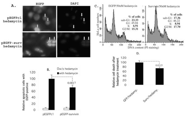
Fig. 8. Exogenous expression of survivin in cancer cells protects cells from apoptosis induced by hedamycin.
A, HeLa cells were transfected with pEGFPc1 control vector and survivin expression vector. Cells were treated overnight with or without hedamycin (50 nM) 24 h after transfection. Cells were then fixed and stained with 4′,6-diamidino-2-phenylindole. Apoptotic cells were evaluated morphologically under a fluorescence microscope. Examples of dead (upper panel) and live (lower panel) cells are shown with arrows. B, the histogram is the relative percentage of cell death under each condition (the EGFP vector-transfected cells with hedamycin treatment were set at 100) based upon the dead cells versus total green cells in four independent microscopic fields. Each bar is the mean ± S.D. C, HeLa cells were transfected and treated as in panel A in 6-well plates. Cells were then processed for flow cytometry. As shown, exogenous expression of survivin decreased sub-G1 DNA contents (dead cells) and accordingly increased S and G2/M cells. D, the histogram shows the relative cell death after hedamycin treatment derived from two independent flow cytometry experiments in triplicate. Each bar is the mean ± S.D. The p values are indicated (B and D).

