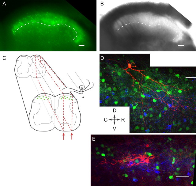
Figure 1.
Spinal cord slice preparation and experimental setup. A, B, The dorsal horn of a transverse P19 spinal cord slice. A, EGFP-positive neurons are visible throughout lamina I–III. B, Under transmitted light, lamina II can be distinguished from other laminae based on its translucent appearance. Medial is on the left, and the ventral border of lamina II is traced with a dotted line in both images (A, B). C, Schematic of parasagittal slices (red dotted line) demonstrating how the attached dorsal root can be preserved and stimulated (arrowhead) while a patch-clamp recording is made from an EGFP-positive GABAergic neuron. D, E, Examples of biocytin-filled EGFP-positive GABAergic neurons (red). The dense band of PKCγ-positive somata and puncta (blue) reveals inner lamina II. Other EGPF-positive neurons are also visible (green). D, A P30 neuron at the lamina I/II border, with processes extending as far ventrally as lamina III. The P35 neuron in E is located more ventrally and its morphology is unlike previously defined subclasses. Arrows indicate Dorsal, Ventral, Rostral and Caudal directions for D and E. Scale bars, 50 μm.

