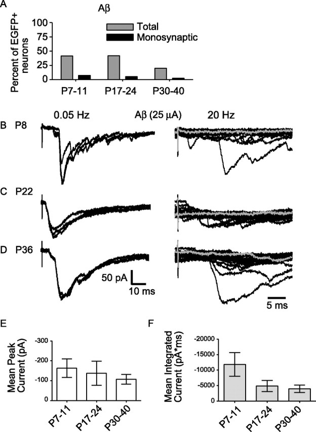
Figure 5.

The prevalence of Aβ fiber drive onto EGFP-labeled GABAergic neurons decreased beyond the third postnatal week, but the strength of Aβ fiber drive was similar during maturation. A, The prevalence of Aβ fiber input to GABAergic neurons at different ages is shown. “Total” includes inputs that are monosynaptic or polysynaptic (gray bars). The prevalence of monosynaptic Aβ fiber input only is also shown (black bars). B–D, Examples of Aβ fiber input in response to 25 μA stimulation recorded from neonatal (P8) (B), juvenile (P22) (C), and mature (P36) (D) GABAergic neurons. On the left are three consecutive traces at low-frequency stimulation (0.05 Hz), and on the right, 20 consecutive responses recorded at high frequency (20 Hz, shown at an expanded timescale). In all three cases (B–D), there are failures at high frequency, consistent with a polysynaptic input (illustrated by gray traces). E, The average peak amplitude of Aβ fiber evoked responses is compared across age groups (17 neonatal, 16 juvenile, and 8 mature neurons; p > 0.05, one-way ANOVA, Newman–Keuls multiple comparison test). F, The average magnitude of Aβ fiber input was measured as mean of the integrated current for responses to 25 μA stimuli at low frequency and compared across all age groups (17 neonatal, 16 juvenile, and 7 mature neurons; p > 0.05, one-way ANOVA, Newman–Keuls multiple comparison test). E, F, Error bars represent SEM.
