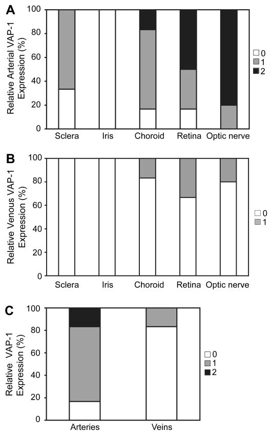
Figure 4. Quantification of VAP-1 Expression in Arteries and Veins in Various Ocular Tissues.
Highest levels of VAP-1 expression were found in the arteries of the retina and optic nerve (A). However, VAP-1 was not detectable in arteries (A) and veins (B) of the iris.
(C) Quantification of VAP-1 expression in the choroidal vessels. VAP-1 expression was significantly higher in arteries than veins.

