
Figure 1. Variability of ocular alignment and motility.
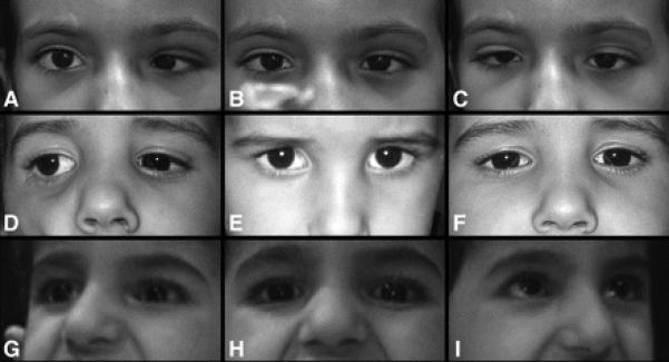
Images A, D, and G are right gaze, while images B, E, and H are in primary position, and images C, F, and I are left gaze. Top row (A, B, and C) are of Patient 4 who had a primary position esotropia with no anomalous head position when fixing with the right eye. This patient was unable to abduct either eye beyond the midline but demonstrated modest adduction bilaterally with globe retraction and fissure narrowing of the adducting eye. Middle row (D, E, and F) are of Patient 8 who was orthotropic in primary position but adopted a small face turn left to maintain comfortable binocular fixation. He had no eye movements to the left but modest symmetric movement of both eyes into right gaze. Bottom row (G, H, and I) are of Patient 5, who was orthotropic in primary position and had severely limited eye movements bilaterally with only minimal adduction the right eye. No abnormal head posture was present in primary position but she used a face turn to view objects to the right or left.