
Figure 3. Variability of MR angiography.
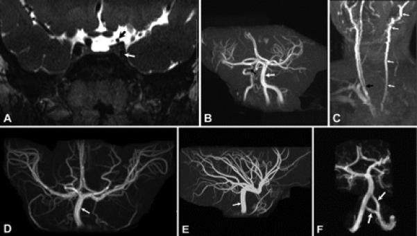
(A) Coronal T2-weighted MR image of Patient 8 demonstrating hypoplasia of the cavernous (white arrow) and supraclinoid (black arrow) portions of the left internal carotid artery. The cavernous and supraclinoid portions of the right internal carotid artery have normal caliber. (B) MR angiogram of the brain of Patient 6 showing absence of the left internal carotid artery and hypoplasia of the cavernous portion of the right internal carotid artery (black arrow). Vertebral and basilar arteries (white arrow) have larger caliber than the right internal carotid artery. (C) MR angiogram of the neck of Patient 6 demonstrating absence of the left internal carotid artery with the common carotid artery ending as a continuation of external carotid artery (white arrows). The right common carotid artery has a bifurcation (black arrow) at about the level of the C7 vertebra, while normal bifurcation of common carotid artery is about the level of C4. (D, E) Anterior (D) and lateral (E) projections of the MR angiogram of the brain of Patient 5 demonstrating absence of both internal carotid arteries and enlargement of the basilar (arrow) and posterior communicating arteries, which supply both the anterior and posterior cerebral circulations. (F) MR angiogram of the brain of Patient 5 showing duplication of the intracranial portion of the left vertebral artery (arrows).