Figure 3.
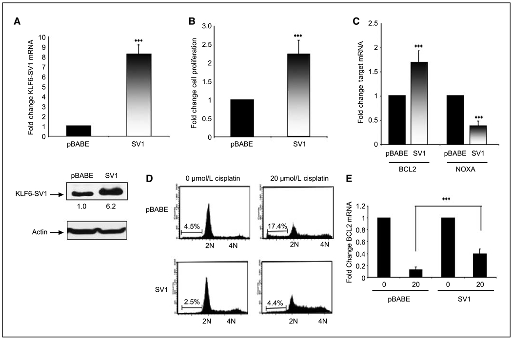
Overexpression of KLF6-SV1 in the A549 lung cancer cell line. A, qtRT-PCR analysis of pBABE and SV1 retrovirally infected A549 cells shows an 8-fold overexpression of SV1 in pBABE-SV1–infected cell lines compared with control cells (pBABE; ***, P < 0.001). B, SV1-overexpressing cell lines proliferate significantly more than control cell lines; tritiated thymidine incorporation was determined at 72 h (***, P < 0.001). Columns, mean change in the rate of cellular proliferation from three independent experiments; bars, SD. C, increased cellular proliferation and survival in SV1-overexpressing cell lines is associated with increased expression Bcl-2 and concomitant decrease in NOXA expression as determined by qRT-PCR (***, P < 0.001). D, overexpression of SV1 abrogated the proapoptotic effects of cisplatin; pBABE and pBABE-SV1 cells were plated at equal densities and treated with 20 µmol/L of cisplatin; 72 h after treatment, cells were harvested. FACS analysis of the treated cell lines reveals a marked reduction in the induction of apoptosis in SV1-overexpressing cells treated with cisplatin (17.4% versus 4.4%; **, P < 0.01). This experiment was repeated three independent times; representative FACS data are shown. E, overexpression of SV1 significantly abrogated cisplatin-induced down-regulation of Bcl-2 expression (**, P < 0.001).

