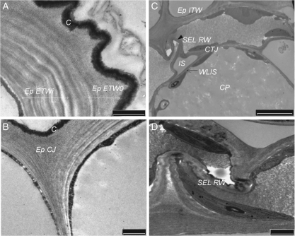
Fig. 2.
Cadmium-induced alterations of the ultrastructure of cell-wall domains within the cortical tissues; PATAg staining. (A) External tangential wall of the epidermis (Ep). Note the tortuous aspect of the tangential wall (ETWo) and of the cuticle (C) and the multilamellate structure of the internal region close to the plasma membrane (ETWi). This figure was observed in 60 % of the sections compared with 10 % slight waving noted in the control sections. (B) Cell junction of the epidermis (Ep CJ). (C) Sub-epidermal layer (SEL) and cortical parenchyma (CP). In cortical parenchyma, note the well-formed intercellular spaces (IS) and the PATAg-contrasted cortical tricellular junction (CTJ). In SEL, the radial walls (RW) appeared highly compressed and Z-shaped (arrow; 100 % sections). (D) Detail of radial wall of sub-epidermal layer (SEL RW). ITW, Internal tangential wall between Ep and SEL; WLIS, wall lining the intercellular spaces. Scale bars: (A, B, D) = 1 µm; (C) = 5 µm.

